

Hip Dysplasia
Hip dysplasia is a condition in which the socket of the hip joint doesn’t fully support the ball of the joint. The condition can create a gradual misalignment or dislocation of the hip, which can wear down cartilage and lead to early-onset osteoarthritis of the hip. A dysplastic hip can also lead to an acetabular labral tear (a torn labrum, which is a soft tissue that lines and secures the hip joint socket).
Although it is sometimes also called "congenital hip dysplasia" or "congenital hip dislocation," the modern and more precise term is "developmental dysplasia of the hip" (or DDH), since the condition is not always present at birth ("congenital") nor does it always involve a dislocated hip. It is usually diagnosed in newborns and young children, although it can develop later and affect teenagers and adults.
Anatomy of the hip
The hip is a ball-and-socket joint composed of two bones connected by cartilage, ligaments, tendons and muscles:
- the femoral head or tip of the femur (thighbone) is the ball
- the acetabulum, a cup on the side of the pelvis, is the socket
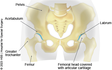
In hip dysplasia, the acetabulum does not develop fully, making it too shallow to adequately contain and support the femoral head. The ball and socket of the hip become misaligned and the hip labrum can end up bearing the forces that should be distributed throughout the hip. Also, more force is placed on a smaller surface of the hip cartilage and bone, resulting in arthritis over a number of years.
Symptoms of hip dysplasia include pain in the groin and/or on the side or back of the hip joint. These symptoms can be distinguished from "growing pains," which are most common in kids under 10. Growing pains in the legs, knees and hips are usually felt at night, after the child has been active during the day, but then go away by the next morning. In a more serious condition like hip dysplasia, the pain will remain constant or increase over time. A child or young adult with hip dysplasia may also hear a sound – usually characterized as clicking, snapping or popping – when moving the hip during activity. A person with hip dysplaisa may also develop a limp to avoid the pain.
There is a wide range of severity among hip dysplasia cases. Milder cases may not be noticed until adolescence or young adulthood. More severe cases can usually be detected during the perinatal period (shortly before or after birth). There are several risk factors that increase the likelihood of hip dysplasia in a prenatal or newborn child. For children who have one or more of these risk factors, it is recommended that an ultrasound be performed at around four to six weeks of age to see whether or not the hip is developing normally.
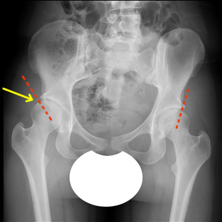
Anterior to posterior (front to back) X-ray showing hip dysplasia in the right hip (at the yellow arrow).
The dotted red lines illustrate the lack of acetabular coverage in the dysplastic hip vs. the healthy hip.
Treatments for Hip Dysplasia
People with hip dysplasia don’t always need surgery. If the condition is diagnosed early (in the prenatal period or during infancy) it can often be treated effectively with bracing. A mild hip dysplasia may not require any treatment, but may need to be monitored as the child grows. In such cases, complications may never arise or they may arise only once the child becomes an adolescent or young adult.
For more serious cases, hip dysplasia in a skeletally immature child can be treated with a variety different surgical procedures of the pelvis and acetabulum. In a skeletally mature teenager or young adult, a procedure called periacetabular osteotomy is done. In a PAO, portions of the pelvis are cut in order to reposition the acetabulum so that it gives better coverage to the femoral head. This surgery has great potential to prevent or delay hip arthritis, especially if it is performed prior to irreversible cartilage injury, such as a torn labrum.
For more information, read the articles on hip dysplasia below.
Explore Related Patient Stories
View All Patient Stories
Hyacinth Bouzalakos
Baldwin, NY
Hip Labral Tear
Melissa Somarriba
Danbury, CT
Hip Labral Tear
Arleen Mitchell
New York, NY
Hip Replacement Surgery

Richard Matthews
Brooklyn, NY
Hip Replacement Surgery

Grace Sobczyk
Butler, NJ
Hip Replacement Surgery

Sierra Lavine
New York, NY

Morgan K
Merrick, NY
Rehabilitation
Amanda Yonks
Hip Labral Tear

Debbie Choo
Mountainside, NJ
Hip Replacement Surgery

Alex Durning
New York, NY

Angela Reynar
Millerton, NY
Hip Replacement Surgery

Colette Cipriano
Colts Neck, NJ
Osteotomy
Killian
Brooklyn, NY
Hip Dislocation (Dislocated Hip)

Charlie G
New York, NY
Rehabilitation
Elana Yoffie
Boston, MA

Caroline Park
Greenwich, CT
Katelyn Vinci
Pearl River, NY
Matthew Reyes
Westbury, NY
Rehabilitation

Marie Pritchett
Trumbull, CT
Rehabilitation

Erin Flood
Medford, NY