

MACI (Matrix-induced Autologous Chondrocyte Implantation)
What is matrix-induced autologous chondrocyte implantation (MACI)?
Matrix-induced autologous chondrocyte implantation, or MACI, is a procedure in which a patient’s own cells are used to regrow new cartilage for the knee joint. MACI has been approved by the U.S. Food and Drug Administration (FDA) since 2016 to treat cartilage damage in the knee in adults. Because the patient’s own cells must first be grown in a lab, it is a two-part procedure.
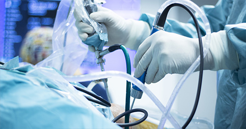
- First, a cartilage biopsy is performed arthroscopically. This allows the surgeon to look inside the knee with a camera, remove a small piece of the patient’s healthy cartilage, and then send it to a lab where the cells are grown.
- When the cells are ready, they are delivered on a thin collagen patch (the MACI implant). The surgeon cuts the implant to fit the defect in the patient’s knee and secures it in place. The cartilage cells then grow and strengthen during recovery. At HSS, this second step can be performed through a small open incision or, for many patients, entirely arthroscopically (arthroscopic MACI).
Who is a candidate for MACI?
MACI candidates include:
- Patients with symptomatic, full-thickness cartilage defects in their knee, including patients with more than multiple defects or those with complex shapes.
- Patients with an unstable kneecap (patellofemoral instability patellofemoral instability) that has caused painful cartilage degeneration in their patellofemoral joint (kneecap or trochlea).
- Patients whose knee is otherwise healthy, without widespread arthritis. Related problems, such as leg malalignment, an unstable kneecap, or a torn ligament or meniscus, can often be corrected at the same time.
Symptoms concerning for cartilage defects include sharp or dull pain in knee that is amplified by activity and weight bearing, recurrent knee swelling, and catching or locking sensations. Patellar instability symptoms include the kneecap coming off its track and subluxing or dislocating.
At HSS, candidates for MACI surgery will be evaluated by their surgeon using both a physical examination as well as magnetic resonance imaging (MRI) to provide the clearest picture of the cartilage injury present in the knee. The size and location of the cartilage damage help the surgeon determine whether MACI, and in particular arthroscopic MACI, is the best option.
What happens during a MACI procedure?
During the first operation, the surgeon views the knee through two small portals. Using a very small camera, the surgeon can examine the condition of the cartilage. If necessary, the surgeon may debride – remove damaged tissue – from the knee, and then will take a biopsy of the patient’s cartilage. This procedure takes around 30 minutes. The biopsy is then sent to the lab, where the cells are grown and placed on a collagen matrix over approximately one month.
During the second operation, the surgeon prepares the area of damaged cartilage and cuts the MACI implant grown in the lab to the exact size of the defect in the patient’s knee. The implant is then secured onto the defect with a surgical glue. The second operation is also where a combined procedure would take place (such as tibial tubercle osteotomy or MPFL reconstruction) if needed. At HSS, the implant can be placed in one of two ways: open MACI and arthroscopic MACI.
Open MACI
The implant is placed through a small incision (about 5 cm or 2 inches) in the front of the knee. This procedure takes approximately one hour. The open approach may be preferred for larger defects, defects in harder-to-reach areas such as the back of the kneecap, or when another open procedure is being performed at the same time.
Arthroscopic MACI
Some HSS surgeons offer arthroscopic MACI, an advanced technique approved by the FDA in 2024. Using a small camera and specially designed instruments, the surgeon prepares the defect and places the implant through small incisions, without the need to open the knee joint. Arthroscopic MACI is approved for cartilage defects up to four square centimeters (4cm2), which is roughly the size of a postage stamp. Smaller incisions mean less disruption of the tissues around the knee, and studies of patients treated with arthroscopic MACI have shown lasting results more than 10 years after surgery.
What is recovery from MACI knee surgery like?
Since MACI is a two-step procedure, there will be two recoveries following each of the stages:
- Stage 1: The patient returns home from the hospital the same day they receive the procedure. The patient can immediately bear weight as tolerated, and most patients are off crutches within a few days. Patients can progress with activity as tolerated. Most patients can return to work within a day or two.
- Stage 2: The patient returns home from the hospital the same day they receive the procedure. Patients will wake up from the surgery in a knee brace and will typically wear the brace for about six weeks. How much weight a patient may put on the leg depends on where the cartilage was repaired. Patients treated for defects of the kneecap or trochlea (the groove the kneecap glides in) are often allowed to bear weight as tolerated with the brace locked straight. Patients treated for defects on the end of the thigh bone (femoral condyle) typically use crutches and gradually increase the amount of weight on the leg, reaching full weight bearing at around 6 to 8 weeks. Patients will also be instructed to use a continuous passive motion machine (CPM) to minimize scar tissue, encourage even cartilage growth, and keep the joint moving without actively using the patient’s muscles. Physical therapy typically begins within the first few weeks after the second operation.
The new cartilage continues to mature for a year or more after surgery. Most patients return to low-impact activities, such as stationary biking and swimming, within the first few months. Return to running and sports is gradual and guided by your surgeon and physical therapist, and many patients return to sports between 10 to 12 months after surgery.
What results can I expect from MACI surgery?
Patients who are good candidates for MACI should expect significant improvement in knee pain and function. Studies that have followed patients for 10 years or longer show that these improvements last: about 9 out of 10 patients are satisfied with their pain relief, and fewer than 1 in 10 go on to need a knee replacement.
Long-term results after arthroscopic MACI are similarly encouraging, with high patient satisfaction more than 10 years after surgery. Goals should be discussed with your surgeon prior to selecting a procedure, but patients are often able to return to sports and activities that their knee pain was preventing them from participating in.
Research from the authors of this article, who specialize in cartilage surgery at the HSS Institute for Cartilage Repair has found that about 80% of athletes return to sport after cartilage restoration surgery, and that MACI is among the procedures most likely to allow athletes to return at the same or a higher level.1 As with any surgery, there are risks that should be discussed with your surgeon before undergoing MACI surgery.
Key takeaways
- Matrix-induced autologous chondrocyte implantation (MACI) uses a patient’s own cartilage cells to repair a cartilage defect. The cells are collected arthroscopically, grown in a laboratory, and later implanted into the cartilage defect, making MACI a two-stage procedure.
- MACI may be appropriate for patients with painful knee cartilage defects or cartilage degeneration associated with patellar instability. A surgeon assesses the injury through a physical examination and MRI, while also considering symptoms such as anterior knee pain or kneecap instability.
- During the first operation, the surgeon takes a cartilage biopsy that is grown on a collagen matrix for approximately one month. In the second operation, the resulting implant is shaped to match the cartilage defect and secured within the knee.
- Weight-bearing after the second MACI operation depends on the location of the cartilage repair. Patients with kneecap or trochlear repairs may bear weight with a locked brace, while those with femoral condyle repairs typically progress more gradually.
- MACI rehabilitation continues as the implanted cartilage matures for a year or longer. Physical therapy begins within the first few weeks, low-impact exercise resumes within several months, and many patients return to sports between 10 and 12 months.
Authors


References
- Kunze KN, Mazzucco M, Thomas Z, Uzzo R, Rodeo SA, Warren RF, Wickiewicz TL, Williams RJ 3rd. High Rate of Return to Sport for Athletes Undergoing Articular Cartilage Restoration Procedures for the Knee: A Systematic Review of Contemporary Studies. Am J Sports Med. 2025 Aug;53(10):2471-2482. https://pubmed.ncbi.nlm.nih.gov/39790040/